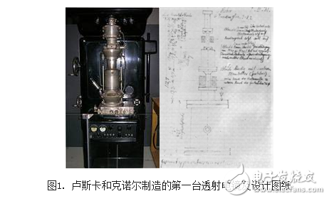
Transmission Electron Microscope (TEM) is a large-scale microscopic analysis device that uses a high-energy electron beam as an illumination source for magnification imaging. In 1933, German scientists Ruska and Knoll developed the world's first TEM (see Figure 1). In 1939, Siemens used this electron microscope as a prototype to mass-produce. The first batch of commercial transmission electron microscopes, about 40 units, has a resolution that is 20 times higher than that of optical microscopes. Since then, human beings have had more powerful weapons for scientific research in the microcosm. Today, TEM has been born for more than 70 years. The electron microscopy, which is formed by the application of electron microscopy, has become more and more perfect. The resolving power of electron microscopy has been improved by more than 100 times than the original, reaching the Asian-European level. And play an increasingly important role in natural science research. 1) Due to the limitations of sample preparation techniques, for most biological samples, generally only 2nm resolution is achieved. 2) The resolving power of an electron microscope image depends not only on the resolution of the electron microscope itself, but also on the contrast of the sample structure. 3) The light source used in the electron microscope is an electron wave, and the wavelength has no color reaction in the non-visible range, and the formed image is a black and white image, and the image must have a certain contrast. 4) The biological tissues and cellular components are mainly composed of light elements such as C\H\O\N. Their atomic number is low, the electron scattering ability is weak, and the difference between them is very small. The contrast of the image under electron microscope is generally smaller. low. 5) Due to the weak penetration of the electron beam, the sample must be made into ultra-thin sections. 6) The observation surface is small, the net can be directly 3mm, and the ultra-thin slice range is 0.3-0.8mm. 7) Strong irradiation of the electron beam, easy to damage the sample, deformation, sublimation, etc., or even breakdown and breakdown, may cause the observation structure to produce artifacts. 8) The electron microscope barrel must be kept vacuum during observation. In order to ensure that the sample is not damaged under vacuum, no moisture should be required for the sample. Therefore, biological samples of living organisms cannot be observed. 9) The biological sample preparation is complicated. In the process of multi-step preparation, the sample is prone to structural changes such as shrinkage, expansion, breakage and loss of inclusion loss. HuiZhou Superpower Technology Co.,Ltd. , https://www.spchargers.com